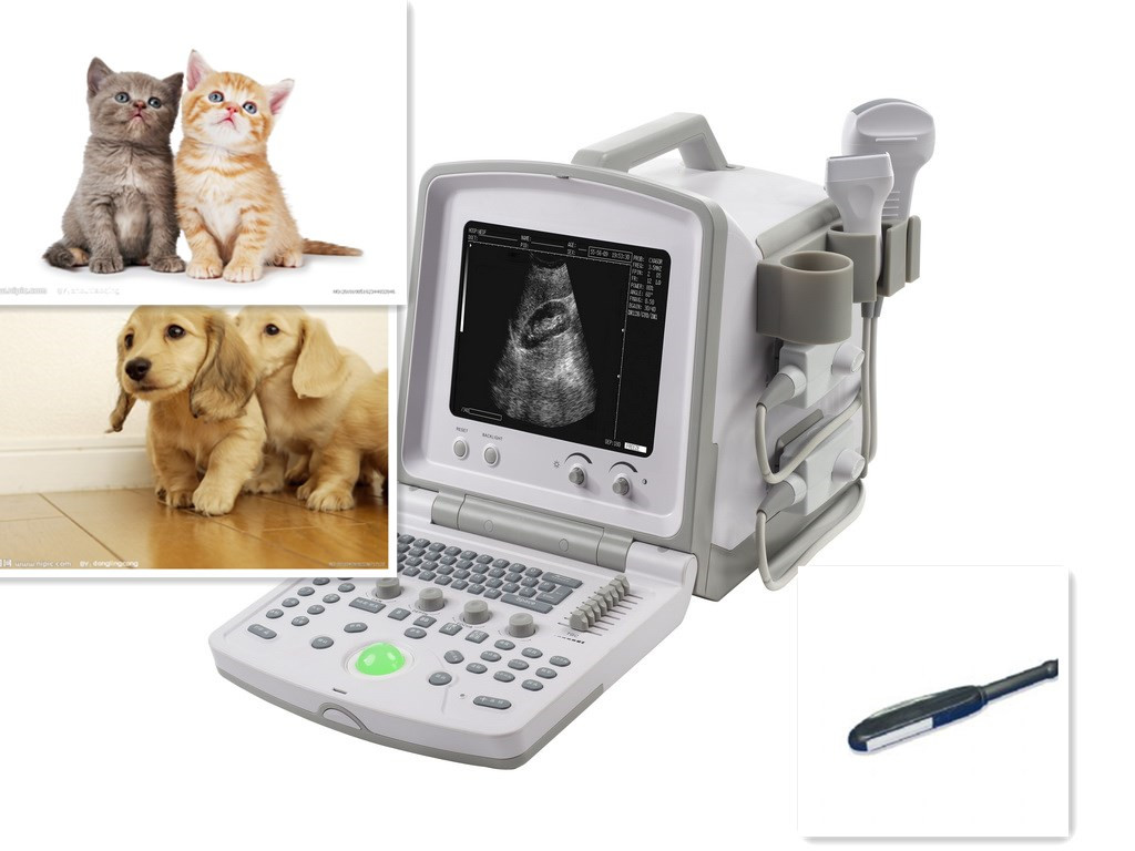
1. Brief Introduction
l This equipment is high resolution linear ultrasound scanning diagnostic equipment. It adopts micro-computer control and digital scan converter (DSC), digital beam-forming (DBF), real time dynamic aperture (RDA), real time dynamic receiving apodization, real time
Dynamic receiving focusing (DRF),
Digital frequency Scan (DFS),
8 segments TGC, frame correlation technologies to endue its image with clarity, stability and high resolution.
l There are six display modes: B, B+B, B+M, B+M/M, M and 4B; And 256 gray scale.
l The system can process real time image display, freeze, save, load, zoom, up and down flip,
left and right flip, black and white flip,
and capacity cine loop; Multi-level scanning depth, angle, dynamic range, acoustic power, frame correlation factor regulation and focus number,
focal space, focus position, etc. It offers more than 16 body marks.
l Date, clock display; Name, sex, age, doctor, hospital annotation; Distance, circumference, area, volume measurement;
EDD Measurement for equine, bovine, sheep, dog and cat. Many probes are optional for clinic diagnosis demands.
l PAL-D video output offers connection to external video image printer and big display and other equipments. High speed
USB port provides real time image transfer to the PC.
l Adoption of folded soft push keyboard and trackball provides immediate, convenient and flexible operation. The equipment
is jet molding enclosure and potable structure, the usage of non-industrial frequency transformer switching power supply, programmable parts (FPGA) and surface mounting technology (SMT)
make the whole unit highly compact.
2. Range Of Application
Suitable for diagnosis on horses, cows, sheep, pigs, cats and dogs and other animals.
3. Technical Specification
|
Probe |
C3-1/60R/3.5 MHz
C5-1/60R/3.5 MHz
convex array |
L3-1/7.5Mhz
HF linear |
C1-6/20R/5.0MHz
micro convex |
LV2-5/6.5MHz endo-rectal probe |
|
Display depth (mm) |
240 (max) |
|
Maximal detect depth (mm) |
≥190 |
≥80 |
≥90 |
≥90 |
|
Resolution
(mm) |
Lateral |
≤2
(depth≤80)
≤3
(80<depth≤130) |
≤1
(depth≤60) |
≤1
(depth≤40) |
≤1
(depth≤60) |
|
Axial |
≤1
(depth≤80) |
≤0.5
(depth≤80) |
≤0.5
(depth≤40) |
≤0.5
(depth≤80) |
|
Blind zone(mm) |
≤3 |
≤3 |
≤2 |
≤3 |
|
Geometric position precision |
Horizontal |
≤0 |
≤3 |
≤4 |
≤3 |
|
Vertical |
≤3 |
≤5 |
≤2 |
≤5 |
|
Monitor size |
10 Inch |
|
Display mode |
B, B+B, B+M, B+M/M, M, 4B |
|
Image gray scale |
256 Scale |
|
Cine loop |
≥500 Frame |
|
Image storage |
64 Frame | <, /TR>
|
Scan angle |
Adjustable |
|
Scan depth |
40mm-240mm |
|
Image flip |
Up/down, left/right, black/ white |
|
Image Process |
GAMA, Image Smoothen, THI, Histogram, Zoom |
|
Focus |
Focus Number, Focus position, Focal space |
|
Measure |
Distance, circumference, area, volume, heart, GA, EDD |
|
Character display |
Date, clock, name, PID, age, Sex, hospital name, doctor, |
|
Notation |
full-screen character editor, posture mark, Position indication |
|
USB port |
USB2.0 |
|
power consumption (MAX) |
100VA |
|
Net weight |
6.4kg |
|
Size |
365mm×291mm×300mm |
4. Standard configurations
ü Mainframe
ü LV2-5/6.5MHz endo-rectal probe
ü PS cable
ü 2 pieces of fuse tube
ü User Manual
ü Final examination report
ü Packing List
5. Optional pieces
ü 5.0MHz Micro-convex probe
ü 7.5Mhz HF linear probe
ü 3.5MHz or 3.5MHz convex array
ü High speed USB cable
ü Monitor |